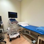
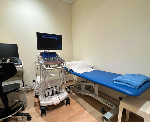
Ultrasound scanning is a diagnostic procedure in which very high frequency sound waves (inaudible to the human ear) are passed through the body. These reflected echoes are detected and analysed to build a picture of your internal organs. This ultrasound scanning procedure is painless and safe. It also gives out real-time information to our medical team.
Ultrasound scanning is used to examine soft organs like the liver, gallbladder and the pelvic region to detect any abnormality.
As a soft tissue imaging technique, its advantages are as follows:
It is non-invasive and does not usually require special patient preparation. It is simple and convenient and easily accepted by most patients including children.
It is safe for the patient and the operator as it does not require ionising radiation. Examinations can be safely repeated as often as the clinical situation demands.
It provides information on structural boundaries as it demonstrates anatomy rather than function. Most organs can be investigated and their shape, size, position and spatial relationship can be determined. It enables tissues to be differentiated on the basis of their interaction with ultrasound scanning.
Cystic structures may be readily distinguishable from solid ones. It can be used to detect and examine moving structures and the pattern of movement can be determined.
The use of ultrasonic doppler technique to study flow patterns in arteries and veins is now well known. It is possible to detect changes in flow speed and direction and thus to evaluate many of the diseases of the arterial and venous system.
Preparation
Before the examination
Different examinations have specific preparation requirements. Please check with your doctor/ nurse if you are unsure.
If you have taken similar examinations in the past, it is advisable to bring them along on your appointment.
When making your appointment please inform the nurse if you are on medications for conditions like asthma, diabetes, cardiac or kidney diseases etc, or if you think you may be pregnant.
If you will be taking a mammogram examination, you may wish to be comfortably attired (e.g. wearing a button-on top) for ease of examination. Do not use powder or deodorant on your breasts or armpits on the day of your examination as it will affect the mammogram image.
During the examination
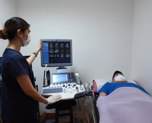
When your appointment is due, you will be led to the examination room by the radiographer.
You will then be requested to position yourself either on the examination bed or to remain standing when the examination is conducted. This will be explained clearly to you by the radiographer who will be conducting the examination. At no time should you be feeling uncomfortable and if you are, do inform the radiographer immediately.
Depending on the type and complexity of the examination, each examination will usually take between 10 – 35 minutes.
After the examination
The collection of results will depend on your doctor’s request. The result will either be collected by you where you will bring it back to your doctor for further consultation or it will be dispatched to your doctor who will then schedule an appointment to discuss the findings with you.
Ultrasound Scanning is a technique which uses high frequency ultrasonic waves to produce images. It is radiation free and safe for everyone including pregnant women and children.